
FDA Approves High-resolution MRI, Better at Spotting Pituitary Tumors in Cushing’s Patients
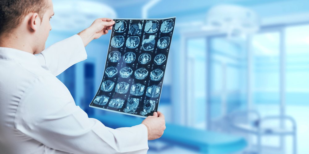


The U.S. Food and Drug Administration has approved the clinical use of a magnetic resonance imaging (MRI) scanner — the ultra-high-field 7T Terra MRI — with unprecedented resolution that allows for more reliable images of the brain.
The approach recently allowed the precise localization of a small tumor in the pituitary gland, which standard MRI had failed to spot, in a patient with Cushing’s disease.
So far, only one scanner of this kind exists in the U.S.. It was installed in February 2017 at the Mark and Mary Stevens Neuroimaging and Informatics Institute (INI) of the Keck School of Medicine, University of Southern California (USC).
The new scanner has an increased magnetic field strength of 7 Tesla, which is more than four times that of conventional MRI. This property greatly improves the instrument’s signal-to-noise ratio, dramatically increasing the spatial resolution and contrast of its images so that scientists can visualize the human living brain in high-definition and with unprecedented detail.
The 7T Terra is ideal for high-resolution neuroimaging, exploration of neurodegenerative diseases such as Alzheimer’s and Parkinson’s, and diagnosis and treatment of other brain diseases, a USC news story by Zara Greenbaum states.
Earlier this year, a report described the case of women with Cushing’s disease with a pituitary adenoma (slow-growing, benign tumor in the pituitary gland) that was possible to localize only with the new 7T MRI.
Based on laboratory analysis that revealed high levels of adrenocorticotropic hormone (ACTH) and cortisol, the doctors suspected a pituitary adenoma and recommended the patient for surgery. However, they ignored the precise location of the tumor, which failed to be detected by standard MRI scanners (1T and 3T).
Two hours before surgery, the woman underwent a 7T MRI scan which finally identified with high precision the location of the adenoma, a very small tumor of 8 mm on the right side of the pituitary gland.
“The 7T may save patients an invasive procedure. It also makes it easier for neurosurgeons to selectively remove a tumor without damaging surrounding areas,” said Gabriel Zada, MD, associate professor of neurological surgery at the Keck School.
Since its arrival, the device has supported exploratory research into into both healthy and diseased brains.
Now the scanner’s advanced imaging technology can be used to help with diagnosis, treatment and monitoring of patients with neurological diseases, including Cushing’s disease.
“This device, which has already made its mark as a powerful tool to advance research in the neurosciences, is now accessible to clinical populations in addition to researchers,” said Arthur W. Toga, PhD, provost professor and chair at the Keck School and director of the USC Stevens INI.
“Clinicians across the university and beyond can now leverage all the benefits of increased spatial resolution to serve patients in need,” he said.