
Cushing’s Linked to Structural, Functional Changes in Brain’s Hippocampus
Written by |


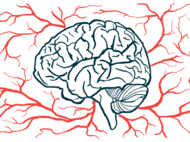
People with Cushing’s disease have structural and functional changes in the hippocampus — a brain region involved in memory, emotions, and stress responses — relative to healthy individuals, a small study shows.
All four hippocampus subregions were smaller in Cushing’s patients and the connectivity to brain networks involved in cognitive function, behavior, and emotion regulation was impaired and associated with quality of life.
The findings illustrate the damaging effects of excessive levels of the stress hormone cortisol — the cause of Cushing’s — in the hippocampus and shed light on what’s behind Cushing’s cognitive and mood symptoms, the researchers noted.
They emphasized the need for effective therapies in stress-related brain conditions to try to prevent further hippocampal damage.
The study, “Altered hippocampal volume and functional connectivity in patients with Cushing’s disease,” was published in Brain and Behavior.

Mental health issues such as anxiety and depression, sleep disturbances, and cognitive impairments are among the many symptoms caused by the abnormally high cortisol levels that mark Cushing’s. Cortisol is a glucocorticoid, or a steroid hormone, produced by the adrenal glands atop the kidneys. How it affects specific brain structures and functions remains largely unclear, however.
The hippocampus, a brain region rich in glucocorticoid receptors and part of the body’s stress response system, is thought to be easily affected by chronic cortisol excess. It’s involved in memory and mood regulation and glucocorticoid injections in the hippocampus of animal models were shown to affect nerve cells’ structures involved in neuronal communication.
Previous studies suggested that “both structural and functional alterations in the hippocampus might contribute to the psychotic symptoms in CD [Cushing’s disease] patients,” the researchers wrote.
A team of researchers in China evaluated the volume and functional connectivity of all four hippocampal subregions (left and right rostral hippocampus and left and right caudal hippocampus) in 47 Cushing’s patients and 53 healthy people who were used as controls.
The rostral hippocampus (located towards the face) is involved in stress, emotion, and affect, while the caudal hippocampus (located towards the neck) performs primarily cognitive functions.
Hippocampus structural and functional changes were assessed with brain MRI. Functional assessments focused on hippocampus connections with the default mode network (DMN), the frontoparietal (FTP) network, and the limbic network due to their role in stress-related psychiatric conditions.
The DMN supports self-related cognitive functions, the FTP is involved in cognitive regulation of behavior and emotion, and the limbic network plays a key role in processing emotion and memory.
All participants had their cortisol levels measured and underwent a comprehensive neuropsychological assessment. Quality of life and neuropsychiatric symptoms were also assessed in Cushing’s patients with validated measures to evaluate potential links with hippocampal changes.
Results showed no significant differences in terms of age, sex, and years of education between the groups, but Cushing’s patients had higher cortisol levels, and scored lower on cognitive measures and higher in anxiety and depression scales.
People with Cushing’s also had significantly smaller volumes of all four hippocampal subregions and altered patterns of hippocampal functional connectivity relative to controls.
Specifically, the right rostral hippocampus showed significantly increased functional connectivity with a DMN region, while both the right and left rostral hippocampus exhibited reduced connectivity with a component of the FTP network.
Significantly altered functional connectivity was found between the left caudal hippocampus and the DMN regions, and the right caudal hippocampus showed lower connectivity with a DMN region and increased connectivity with some limbic regions.
Moreover, higher values of functional connectivity between the left caudal hippocampus and a DMN region called the anterior cingulate cortex were significantly associated with better quality of life scores in Cushing’s patients. No other links were found between structural or functional changes and quality of life or neuropsychiatric symptoms.
These findings “elucidate the cumulative effect of cortisol on the [structure] and function of hippocampus and provide important information to further understand the role of hippocampus in stress-related brain disease,” the researchers wrote.
Given that “emerging evidence proposes that interactions within and between these large-scale brain networks play important roles on brain functions and may be affected in multiple psychiatric disorders,” excess cortisol may disrupt hippocampal interactions with these regions and increase “vulnerability for emotional and cognitive problems,” they wrote. “In line with this view is evidence that altered hippocampal functional connectivity is associated with the quality of life in CD patients.”
They said the results also “reinforce the need for effective therapeutic interventions in stress-related brain disease to halt possible hippocampal damage,” and noted that larger studies are needed to confirm the findings, along with studies to evaluate changes in hippocampal functional connectivity after cortisol levels normalize with therapy.


Leave a comment
Fill in the required fields to post. Your email address will not be published.