
Fluorescent Dye Significantly Aids Surgeons in Laparoscopic Adrenalectomy


A fluorescent dye that helps surgeons accurately identify anatomical structures is safe and may help guide laparoscopic procedures to remove the adrenal glands, a new study shows.
The study, “Role of indo-cyanine green (ICG) fluorescence in laparoscopic adrenalectomy: a retrospective review of 55 Cases,” which included several Cushing’s disease patients, was published in the journal Surgical Endoscopy.
Laparoscopic adrenalectomy is the gold standard treatment for removal of benign masses in the adrenal glands. The procedure involves the use of a laparoscope, which is a small tool with a camera and a light that allows physicians to see the inside of the body.
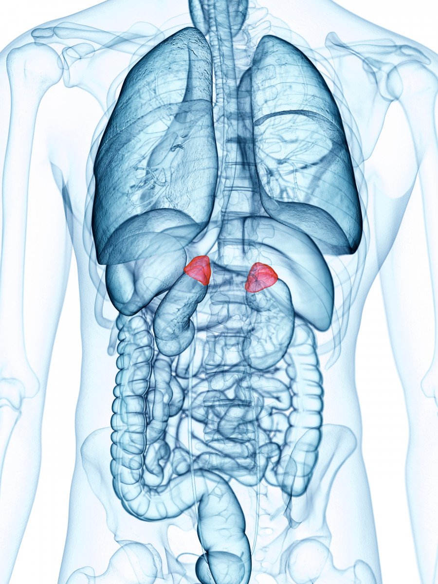
When undergoing such procedures, it is important for surgeons to accurately distinguish the adrenal gland from the surrounding fat.
Indo-Cyanine Green (ICG) fluorescence is an emerging technology that is being used frequently to help surgeons identify the adrenal gland. ICG is a sterile, water-soluble dye that, when injected intravenously, remains largely confined to the blood vessels.
The adrenal gland is surrounded by multiple layers of blood vessels. As such, it has rapid uptake of the ICG dye compared to the surrounding fat tissue.
ICG fluorescence relies on the use of near-infrared (NIR) light, which usually is shed by the laparoscope. A camera with the required filters then can help display the tissues.
One of the benefits of using ICG is that it is eliminated quickly by the liver, which limits potential toxicity.
Researchers in India conducted a study to evaluate the role of ICG fluorescence in laparoscopic adrenalectomy. Specifically, they looked at the benefits and safety concerns associated with the use of this technique.
The study included 55 patients who underwent laparoscopic adrenalectomy using NIR fluorescence-enabled laparoscope and were analyzed retrospectively.
All patients were administered the dye before undergoing surgery. Among them, 54 patients underwent successful laparoscopy using ICG fluorescence. None of the patients died from the surgery, and there were no adverse effects found that were attributed to the use of the dye.
The final diagnoses of the patients included a range of adrenal masses, including adrenocortical adenoma, adrenocortical carcinoma, and Cushing’s adenoma.
“Within our series of 55 cases, all three operating surgeons unanimously agreed that ICG fluorescence routinely helped improve the course of surgery,” investigators wrote.
In particular, the procedure was of significant benefit in patients with smaller glands, where ICG helped differentiate between the adrenal gland and surrounding fat, allowing physicians to start the dissection.
One of the caveats to this method is that the fluorescence usually subsides within 15 minutes. The short duration of fluorescence means that a single dose of ICG is not sufficient for the entirety of the operation.
“The surgical team acknowledges the utility of the technology in real-time differentiation of tissues and identification of vascular structures, providing immediate guidance during surgery,” the physicians concluded. “NIR fluorescence is a safe, useful addition in laparoscopic adrenalectomy which will undergo further refinement over time.”