Degree of Fat in Bone Marrow Ably Judges Fracture Risk in Cushing’s Patients, Study Suggests
Written by |


Measures of the degree of fat in bone marrow could be a reliable marker of bone health, helping doctors to predict the risk of fractures in people with Cushing’s syndrome, a study has found.
The study, “High bone marrow fat in patients with Cushing’s syndrome and vertebral fractures” was published in the journal Endocrine.
Cushing’s syndrome frequently causes bones to become thinner and brittle as a consequence of prolonged exposure to excess levels of cortisol (hypercortisolism), a hallmark of the disease.
Cortisol is one of the body’s stress hormones, with a wide range of effects throughout the body. It acts on bone by blocking calcium absorption, which decreases bone growth and reduces bone mineral density (BMD).
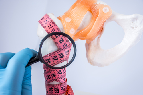
People with Cushing’s syndrome often have porous and weak bones (osteoporosis) that are more vulnerable to breaking, particularly in the vertebrae (the 33 interlocking bones of the spine) and ribs.
Evaluation of bone fragility in these patients is a clinical challenge, because the exam commonly used to measure bone density and diagnose osteoporosis — dual-energy X-ray absorptiometry, or DXA — cannot capture the small abnormalities in bone structure induced by cortisol excess.
In fact, many Cushing’s patients have bone fractures with BMD values that are only slightly low or normal.
A team of researchers with University Hospital AOU Policlinico G. Martino and the University of Messina, in Italy, decided to test a different surrogate of bone fragility in Cushing’s patients — bone marrow fat (BMF).
Marrow fat can be visualized by computed tomography (CT) or magnetic resonance imaging (MRI), and has been proposed as a marker of bone mass, where greater amount of fat indicates lesser bone density and poorer bone health.
Increased BMF has been reported in Cushing’s patients, but it is still unknown whether this variable could be used as reliable marker of bone quality and fracture risk in this population.
The researchers measured lumbar spine BMF in 20 people with active Cushing’s (mean age, 44) and 15 healthy subjects of similar age using magnetic resonance spectroscopy (MRS) and compared those values relative to bone mineral density, and the presence of vertebral fractures.
MRS is a non-invasive exam, conducted on the same machine as conventional MRI, that detects changes in the chemical composition of tissues.
In 16 patients, the disease was dependent on the production of adrenocorticotropic hormone (ACTH), which can be caused by tumors in the pituitary gland (Cushing’s disease) or elsewhere.
Results showed that BMF was significantly higher in Cushing’s patients compared to controls: 52% vs. 27% of fat content, expressed as a percentage of total marrow.
Higher marrow fat was associated with more advanced age, greater levels of midnight cortisol in the blood, and increased serum CTX, a marker of bone resorption or breakdown.
Noteworthy, patients with vertebral fractures — 13 cases, seven of them with severe or multiple fractures — had more fat in their marrow (65% of total marrow) compared to patients without fractures (24%).
Among people with fractures, higher BMF values co-existed with normal or somewhat low bone mineral density measures, more indicative of osteopenia than osteoporosis, and were comparable to measures of more severe bone loss (osteoporosis) or reduced bone mass for their age.
If researchers looked only at patients with normal or low-normal BMD, a higher BMF still associated with vertebral fractures.
This suggests that measuring marrow fat by MRS could be more informative than DXA for identifying Cushing’s patients at high risk of fractures. Magnetic resonance spectroscopy could detect bone fragility in patients that would go otherwise undiagnosed by DXA.
“This study provides a first evidence that vertebral adiposity [fat] may be a marker of hypercortisolism-induced skeletal fragility and measurement of spine BMF could have a role in the diagnostic work-up for the assessment of fracture risk in CS [Cushing’s syndrome],” they wrote.
The scientists believe a link between high BMF and bone fractures related to excessive cortisol exists, because stem cells within the marrow are being converted to fat, rather than to bone-building cells (osteoblasts).