Heart issues seen on MRI despite treatment for Cushing’s: Study
Poorer patient heart function tied to prolonged exposure to high cortisol
Written by |
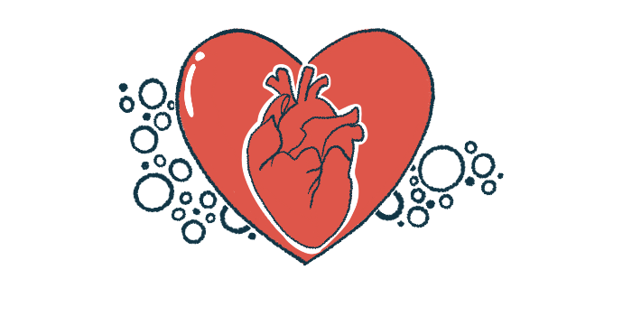


People with Cushing’s syndrome, including those with Cushing’s disease, appear to have more heart issues — like poorer heart function and larger ventricles, which are the heart’s lower chambers — compared with other patients with similar cardiovascular risk factors, a study using MRI imaging has found.
The heart issues in Cushing’s were found despite ongoing treatment for some patients, as well as among others in whom the condition had been cured, according to researchers.
The changes seen on the MRI scans were related to prolonged exposure to high cortisol levels, a hallmark of the condition. While keeping cortisol levels under control may ease heart issues, the changes may remain “persistent and clinically silent,” the team noted, calling for regular monitoring with heart MRI.
The study, “Cardiac magnetic resonance reveals biventricular impairment in Cushing’s syndrome: a multicentre case-control study,” was published in the journal Endocrine.

More heart issues seen in Cushing’s than in patients with other risk factors
Cushing’s syndrome is a group of diseases caused by hypercortisolism, or high cortisol levels. Cortisol is a hormone produced by the adrenal glands located atop the kidneys. In Cushing’s disease, hypercortisolism occurs due to a tumor in the brain’s pituitary gland that ultimately triggers the overproduction of cortisol.
Prolonged exposure to high cortisol levels can increase the risk of cardiovascular problems, such as blood clots, heart attack, stroke, or other issues. Many people with Cushing’s syndrome show abnormalities in the heart’s structure, including hypertrophy, which occurs when the heart muscle becomes abnormally thick.
Now, researchers in Italy sought to determine if such changes persist after cortisol levels are brought under control. To that end, they used cardiac MRI, a noninvasive imaging technique that allows doctors to look in detail into the heart’s structure and function.
The study compared 16 people with Cushing’s syndrome with 15 patients of the same age and sex who had a nonfunctioning adrenal incidentaloma, or NFAI, which is an adrenal tumor that does not release any hormones but is linked to an increased risk of cardiovascular problems. The Cushing’s patients had a mean age of 47.5 years.
Among the Cushing’s syndrome patients, five had active disease but were being treated. The other 11 were cured and manifested no symptoms of the disease. Overall, 12 had Cushing’s disease, whereas four had an adrenal tumor that released cortisol.
Heart MRI scans revealed that patients being treated for Cushing’s had significantly higher systolic and diastolic blood pressure than those who had been cured. Systolic blood pressure is that exerted on arteries when the heart beats, whereas diastolic blood pressure is the one exerted when the heart rests between beats.
The fact that [heart] alterations occur rapidly in [Cushing’s syndrome] and are partially irreversible after remission advocates the use of [heart MRI] to improve the management of fatal cardiac complications.
The researchers then compared patients with Cushing’s syndrome with those with NFAI. They found that the heart’s ventricles — both the left and the right — tended to hold more blood after they contracted in Cushing’s patients than in those with NFAI. More blood left behind suggests weaker heart function.
Consistent with this finding, the ejection fraction of both left and right ventricles, a measure of the percentage of blood that the ventricles pump out with each heartbeat, was lower in people with Cushing’s syndrome than in those with NFAI, suggesting a less efficient heart.
While blood sugar levels tended to be lower in people with Cushing’s, there were no differences in blood pressure or in the levels of fatty molecules in the blood between the two groups of patients. Thus, heart changes were “likely attributable to chronic exposure to cortisol excess independently of known traditional risk factors,” the researchers wrote.
“The fact that such alterations occur rapidly in [Cushing’s syndrome] and are partially irreversible after remission advocates the use of [heart MRI] to improve the management of fatal cardiac complications,” the team wrote.